What is it for?
A Coronary CTA is a heart-imaging test currently undergoing rapid development and evaluation for non-invasively determining whether either fatty deposits or calcium deposits have built up in the coronary arteries, which supply blood to the heart muscle. If left untreated, these areas of build-up, called plaques, can cause heart muscle disease. Heart muscle disease, in turn, can lead to fatigue, shortness of breath, chest pain and/or heart attack.
How does it work?
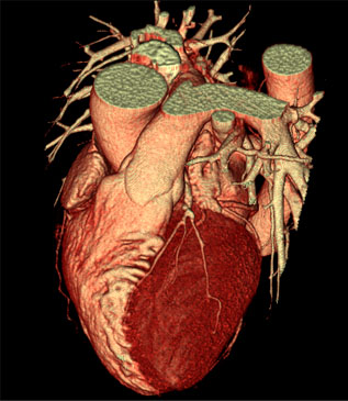
A Coronary CTA comes from a special type of X-ray examination. Patients undergoing a Coronary CTA scan receive an iodine-containing contrast dye as an IV solution to ensure the best images possible. The same IV in the arm may be used to give a medication to slow or stabilize the patient’s heart rate for better imaging results. During the examination, which usually takes about 10 minutes, X-rays pass through the body and are picked up by special detectors in the scanner. Typically, higher numbers (especially 16 or more) of these detectors result in clearer final images. For that reason, Coronary CTA often is referred to as “multi-detector” or “multi-slice” CT scanning. The information collected during the Coronary CTA examination is used to identify the coronary arteries and, if present, plaques in their walls with the creation of 3D images on a computer screen.
How is Coronary CTA different from other heart tests?
One of the most common heart tests is the coronary angiogram, or cardiac catheterization. This test is more invasive and requires more patient recovery time than Coronary CTA. Patients who receive coronary angiograms must have a catheter, or small transport tube, threaded into their coronary arteries, which run along the outside of the heart. The catheter typically is inserted into a blood vessel in the upper thigh and then maneuvered up to the coronary arteries. The catheter then is used to inject the iodine dye needed for the test, which uses X-rays to record “movies” of interior of the coronary arteries.
Although Coronary CTA examinations are growing in use, coronary angiograms remain the “gold standard” for detecting coronary artery stenosis, which is a significant narrowing of an artery that could require catheter-based intervention (such as stenting) or surgery (such as bypassing). On the other hand, this new technology has consistently shown the ability to rule out significant narrowing of the major coronary arteries and can non-invasively detect “soft plaque,” or fatty matter, in their walls that has not yet hardened but that may lead to future problems without lifestyle changes or medical treatment.
Who should consider Coronary CTA?
The single most important step for patients trying to determine whether they should consider a Coronary CTA is consultation with their primary physician. This is because some Coronary CTA uses are more appropriate than others, and the scan carries some risk from X-ray exposure (potential for stimulating cancer) and contrast dye exposure (allergic reactions and kidney damage). Applying careful patient selection and risk-reduction efforts, The Cleveland Clinic has successfully performed more than 13,000 clinical cardiac CT examinations over the past two-year period, many for Coronary CTA.
Overall, Coronary CTA examinations have tended to help determine a lack of significant narrowing and calcium deposits in the coronary arteries, as well as a presence of fatty deposits. This has been found to be particularly valuable in asymptomatic patients with higher risk for coronary disease, in patients with atypical symptoms but lower risk of coronary disease, or in patients with unclear stress-test results. As a result, the Center for Integrated Non-Invasive Cardiovascular Imaging at The Cleveland Clinic currently supports the careful use of Coronary CTA for patients who have:
- Intermediate to high-risk profiles for coronary artery disease, but who do not have typical symptoms (especially chest pain, shortness of breath, or fatigue during heavy physical activity.)
- Unusual symptoms for coronary artery disease (such as chest pain unrelated to physical exertion), but low to intermediate risk profiles for coronary artery disease.
- Unclear or inconclusive stress-test (treadmill test) results.
For these types of patients, Coronary CTA can provide important insights to their primary physician into the extent and nature of plaque formation with or without any narrowing of the coronary arteries. Coronary CTA also can non-invasively exclude narrowing of the arteries as the cause of chest discomfort and detect other possible causes of symptoms. But again, initial consultation with their primary physician is key for patients seeking to determine the appropriateness of Coronary CTA.
Who should not have Coronary CTA?
To date, Coronary CTA has not been proven as effective as the coronary angiogram in detecting disease in the smaller heart arteries that branch off the major coronary arteries. For that reason, Cleveland Clinic physicians do not consider Coronary CTA as an adequate substitute for needed coronary angiography in patients with strong evidence of narrowing of the coronary arteries. Such patients include those with a history of chest pain during heavy physical activity, a history of positive stress-test results, or a known history of coronary artery disease or heart attack. Coronary CTA also is of limited use in patients with extensive areas of old calcified, or hardened, plaque, which is often the case in older patients. Patients who are extremely overweight or who have abnormal heart rhythms also tend not to be suitable candidates for this test because imaging quality is compromised.
Cardiac Computed Tomography or Coronary CT Angiography (MSCT, CT, cardiac CT, coronary CTA or cardiac CAT scan)
Definition:
A traditional CT scan is an x-ray procedure that combines many x-ray images with the aid of a computer to generate cross-sectional views of the body. Cardiac CT uses the advanced CT technology with intravenous (IV) contrast (dye) to visualize your cardiac anatomy, coronary circulation and great vessels. The Cleveland Clinic Foundation uses state-of-the-art multi-row detector CT scanners. With multi-slice scanning, it is possible to acquire high-resolution three-dimensional images of the moving heart and great vessels.
Your doctor uses the cardiac CT to evaluate:
- the heart muscle
- the coronary arteries
- the pulmonary veins
- the thoracic and abdominal aorta
- the sac around the heart (pericardium)
How to prepare for your CTA exam
- Avoid any caffeinated drinks on the day before or the day of your exam. Coffee, tea, energy drinks, or caffeinated sodas.
- Avoid energy or diet pills on the day before or the day of your exam (ask your doctor if you have questions).
- Do not use Viagra or any similar medication on the day before or the day of the exam. It is not compatible with the medications you will receive during the procedure (ask your doctor if you have questions).
- On the day of your exam,do not eat for four hours prior to your scheduled appointment. You may drink water.
- If you are diabetic, ask your physician how to adjust your medications the day of your test. If you think your blood sugar is low, tell the technologist immediately.
- Tell your technologist and your doctor if you are:
- pregnant
- allergic to iodine and/or shellfish or any medications
- undergoing radiation therapy
- over 60 years old or have a history of kidney problems (you may be required to have a blood test to evaluate your kidney function prior to receiving any contrast agent)
What to expect:
- You will change into a hospital gown.
- A technologist will insert an IV line into a vein in your arm to administer contrast (dye) during your procedure.
- You will lie on a special scanning table.
- The technologist will clean three small areas of your chest and place small, sticky electrode patches on these areas. Men may expect to have their chest partially shaved to help the electrodes stick. The electrodes are attached to an electrocardiograph (ECG) monitor, which charts your heart’s electrical activity during the test.
- You will lie on the scanner table, and you will be asked to raise your arms over your head for the duration of the exam.
- During the scan, you will feel the table move inside a donut-shaped scanner. You will receive a contrast agent through your IV to help produce the images. It is common to feel a warm sensation as the contrast circulates through your body.
- Once the technologist is sure that all the information is collected, the IV will be removed.
The CT scan takes about 15 minutes.
After the procedure:
- You may continue all normal activities and eat as usual after the test.
- Your physician will discuss the results of your test with you.
Please ask your doctor if you have any questions about the cardiac CT.
A note about CT and risk:
A CT scan is a low risk procedure. Occasionally, patients experience an adverse reaction to the contrast agent. Some patients develop itching or a rash following the injection. These symptoms are usually self-limiting and resolve without further treatment. Antihistamines can be administered if needed for symptomatic relief. Rarely, a more serious allergic reaction, called an anaphylactic reaction, occurs that may result in breathing difficulty. This reaction is potentially life-threatening and would require medications and treatment to reverse the symptoms. CT scanners use x-rays. For your safety, the amount of radiation exposure is kept to a minimum. Because x-rays can harm a developing fetus, however, this procedure is not recommended if you are pregnant.